There are various schools of thought and practice when it comes to the location of the spinal reflexes on the feet.
To answer this question appropriately we must view the feet as a microcosm (mini-map) of the body. It is popularly agreed by Reflexologists around the world that each foot represents one half of the body. The right foot is made up of reflexes that have connections to the right side of the body, and the left foot is comprised of reflexes corresponding to the left side of the body– the arch of each foot is the body’s dividing line and share the spinal reflexes. The question posed by many is- where exactly is the path of these spinal reflexes on the foot?
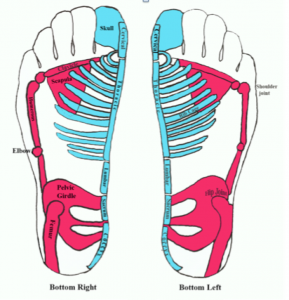
My drawing of the skeletal system as a microcosm within the feet.
Some reflexology practitioners believe the spine can be reflexively stimulated on the edge of the inside of the foot, beginning at the heel bone and following the contour of the arch of the foot to the side of the great toe (as shown in purple in illustration A.) Another point of view is a path just beneath the medial ankle which also follows the contours of the arch to the side of the great toe (as shown in purple in illustration B.) I believe both are valid, and I incorporate each of these approaches into my reflexology sessions– however, I associate the first mentioned reflexive path to the skeletal aspect of the spine, including the appendage of the pelvic girdle attachment, and the later, illustration B, as a reflexive path that innervates the spinal chord and the deeper aspects of the central & peripheral nervous system.
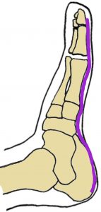
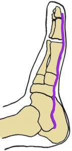
A B
The left foot profile labeled A shows the spinal reflex path I associate with the bones of the vertebral spine which includes the mid-line of the pelvic girdle (the heel.) The foot profile labeled B shows another path for the spinal reflexes I associate with the central nervous system (the spinal chord) that starts along the medial talus.
If we examine how the reflexes of the arch communicates to the spine when we walk, the later, image B, reveals when the spine becomes activated. Follow along with me as I explain how the spine responds during these six stages of walking in one step:
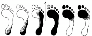
Illustration from Functional Foot Disorders, by Dr. John Martin Hiss
In a healthy stride, the darkened areas show six stages of weight bearing on the foot in one step. Beginning from left to right, the first image exhibits weight placed on the heel of the foot and is then directed to the outer side of the foot. The 2nd stage of walking shows weight continuing along the outer side of the foot toward the toes.
At the 3rd stage of weight bearing, in this one step, weight begins to transfer from the outer foot to the inner foot, via the transverse arch (below)–
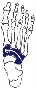
and it is at this point when the medial arch (spinal reflexes) engages, and springs into motion.
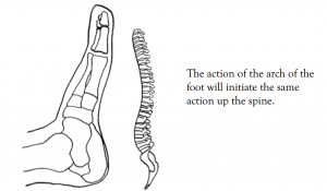
In response to receiving weight, the spring and shock absorbing functions of the arch kick into action. This motion, in-turn, sends a wave of movement throughout the arch of the foot, activating each incremental part of the spine of the body to respond in the like. Ultimately, the central and peripheral nervous system are stimulated both reflexively, and directly, by the locomotion the arch of the foot makes.
The vertical arrow shown in the middle of the six stages of weight bearing image above indicates the time when weight is transferred entirely from the outer column of the foot to the medial column (the arch), which stays suspended in space by the strength of the transverse arch as weight is distributed to the toes. Keep in mind the arch of the foot does not and should not bear weight. Weight gets distributed to the toes by the strong, balancing action of the muscles that attach to the arch.
So with this six stages of walking perspective, the spinal reflexes located just beneath the inner ankle (at the talus) become innervated when the medial column of the foot engages during the 3rd stage of walking. Notice how the darkened area traces the lower spinal reflex at this stage which essentially verifies the path of the spinal reflexes in image B. The inner side of the heel bone (calcaneus) that is traditionally known as the tail bone reflex, would then be reflexive to the inner pelvic bones.
Most importantly, when practicing reflexology, both paths are very effective ways to stimulate the spinal reflexes which serve to relax and enhance appropriate neural connections from the glands to the organ systems of the body, as well as to relieve structural stressors in the feet.
Click here to learn more on how to take care of your feet and spine.
Archives
- October 2024
- December 2023
- October 2023
- March 2022
- July 2021
- August 2020
- May 2020
- April 2020
- February 2020
- January 2020
- December 2019
- November 2019
- October 2019
- August 2019
- June 2019
- May 2019
- April 2019
- January 2019
- December 2018
- October 2018
- September 2018
- July 2018
- June 2018
- April 2018
- February 2018
- December 2017
- November 2017
- October 2017
- September 2017
- August 2017
- July 2017
- June 2017
- May 2017
- April 2017
- February 2017


3 Responses to Where are the spinal reflexes located on the feet?