The late Dr. Simon Wikler, chiropodist and inventor of the shoe for Buster Brown devoted his life to educating the public about the importance of wearing shoes that fit and support the shape and function of feet. The greatest impetus for his passion began after he attributed the early death of his mother to shoes she wore.
After listening to Dr. Wikler talk, I was astounded to hear him say “Locked hips are the kiss of death”. With raised eyebrows I began a quest to get to the bottom of his proclamation.
How do hips get locked? After years of research I was able to substantiate this attrition Dr. Wikler spoke of by linking inhibiting factors that prevent feet from performing their fundamental functions to mostly, like Dr. Wikler suspected, certain shoes and inserts.
It was prominent foot expert, Dr. John Martin Hiss, O.D. who outlined these seven fundamental functions of feet in his book *Functional Foot Disorders:
1. Support: pressure of bones and ligaments against superimposed body weight
2. Balance-control of body weight over the center of gravity
3. Locomotion-coordination of muscles to move joints for propelling the body through space
4. Adaptability-coordination of support, locomotion, and balance to compensate for changed position
5. Distribution-control of contact pressure made by the sole of the foot on the ground as the load moves through the foot during locomotion
6. Vitality-LIFE which manifests itself in the feet as well as the rest of the body. Vitality is the body’s innate intelligence which sustains metabolism
7. Power-refers to the action of muscles that transform chemical energy into mechanical energy
*Functional Foot Disorders, by John Martin Hiss (3rd Edition),Oxford Press, 1949
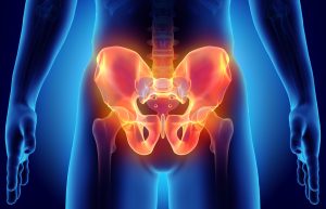
Relating the function of the talus bone to the hips and pelvis
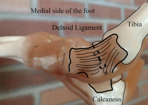
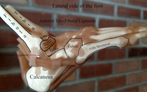
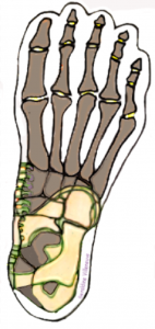
Years of observation unveiled a reflexive correlation with the disruption of any one of these fundamental foot functions to arthritic joints in other parts of the body. For example, the talus, held in place by ligaments that knit the lower leg bones to the foot, is a freely moveable foot bone located beneath the tibia and above the calcaneus. Considered an important part of the true ankle, the talus functions as a hinge joint with the tibia and fibula, however the talus itself has an essential function which is to pivot 45 degrees during locomotion to assist weight to the calcaneus and along the lateral column of foot bones (cuboid, 4th & 5th metatarsals and phalanges.) The ability of the talus to pivot within the ankle has a mutual relationship with the pivotal movement of the ball and socket of the hip. If the talus is not able to pivot because of excessive joint tension it will hinder the dynamic transmission of movement to the hips and pelvis. Diminished hip movement will also decrease the benefit of movement supplied to the vertebral spine which houses the life-line of the body.
In my experience working with feet, consistent variables show when the talus bone appears to be locked amid the tibia and calcaneus. Limited passive and active foot flexion is usually followed by complaints of hip, low-back pain and in many cases groin, and reproductive issues. Based on these findings I find it interesting to note the talus is indeed the reflex area associated with the pelvis, leading me to also conclude once the hips lock, the pelvis and spine may eventually too, and vitality emitted from the spinal cord is likely to lose efficient, neural connections which speaks to Dr. Wikler’s “kiss of death” statement.
Prevention and correction is possible with Structural Reflexology® techniques that release overly tight muscles that move the foot as well as heal hidden ligament injury surrounding the talus to improve ankle and hip movement.
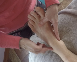
Chronic foot problems persist because of hidden injury in ligaments beneath. In order to improve talo-calcaneal alignment and its hinging function, one must identify and release tension in key ligaments that keep the talus secure. Here’s one way to do this:
Using your index finger and one at a time, gently massage the Deltoid ligaments and the the most commonly sprained ligament, the Anterior Talo-Fibular ligament (ATFL) by crossing their fiber direction. For instance finger direction on the deltoid ligament will move horizontally (left to right). The finger direction on the ATFL will move vertically. Movement should be in one direction only, either forward or backward for approximately 1 minute. Pressure should be firm but not beyond the pain tolerance of the individual you are working with (a 5-6 on a pain scale of 10). After 1 minute check in to find out if the level of pain is decreasing and if so continue for 30 seconds more.